About Color Doppler
Color Doppler is a method of visually detecting motion or blood flow using a color map that is incorporated into a standard B-mode image. The principles of color Doppler are similar to those of pulsed-wave Doppler. However, a larger region can be interrogated, and detected blood flow is assigned a color, typically blue or red, depending on whether the flow is moving toward or away from the transducer. Frequency shifts are estimated at each point at which motion is detected within an interrogated region, thus yielding information on direction of motion and velocity. Shades of blue or red are used to reflect the relative velocities of the blood flow. All stationary objects are represented on a gray scale, as in B-mode imaging. The benefit of color Doppler is that information on the direction and relative velocity of blood flow can be obtained. Color Doppler is limited by its dependence on the relative angle of the transducer to the blood flow.
What is color Doppler test in pregnancy?
Doppler ultrasound uses sound waves to detect the movement of blood in vessels. It is used in pregnancy to study blood circulation in the baby, uterus and placenta . Using it in high- risk pregnancies, where there is concern about the baby's condition, shows benefits
Diagnosis is achieved by performing a Doppler ultrasound of the legs. The test itself takes 15-30 minutes to perform. The timing of the result depends on who interprets the findings (radiology technician, radiologist, vascular medicine physician, etc).
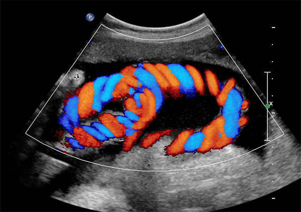
Colour Doppler permits non-invasive assessment of morphology and function of thoracic disease, as reflected in the organ blood supply and perfusion. Vascular structures in pulmonary consolidation are visualised as thin-walled, echo-poor pulsating, branched tubular structures arising from the hilar region, with blood flow detected by Doppler.